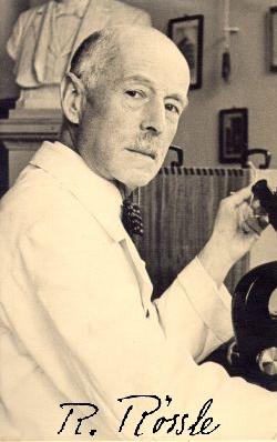
Robert Rössle has been endowed with and has set an example to the virtue of superior professorship (Doerr 1976):
Newton in science, Goethe in teaching!
His pioneering work dealt with introducing objective standards and the quantification of features that are relevant for the description and diagnostic judgement of organisms, organs, tissues and even cells. He was father of the scientific investigation of processes involved in inflammation and allergy.
Maß und Zahl in der Pathologie (measure and number in pathology) together with Frédérick Roulet, Berlin 1932.
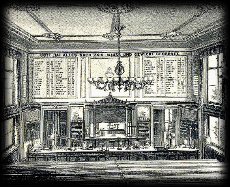
God did arrange everything as to number, measure and weight. Institute of Chemistry in Leipzig at about 1864: the front wall of the main lecture room (Quadbeck-Seeger 2007):
philosophy of this web presentation
This publication will be dealing with special topics of cytometric interest in more detail, which might be helpful to understand, why cytometry sometimes works but often fails.
Nevertheless, the old experience is valid still today:
Each failure has its reasons, each success its mystery.
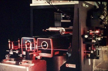
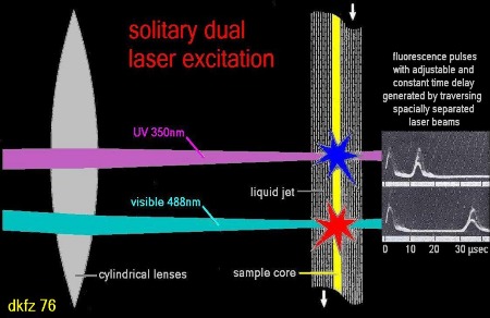
historical remark on multi laser excitation in flow cytometry
multi
laser
flow
cytometry
in
the
beginning
The first cell sorter, equipped with a second laser, was established in 1975. With spatially separated laser foci and solitary (time delayed) excitation two colour fluorescence analysis could be performed using only one detec- tor.
This instrument has been applied with various combinations of fluorescent dyes. For the first time, the profit of Hoechst 33258 and DAPI - SR101 for cell cycle analyses of fixed cells and the Hoechst derivative 33662 to- gether with PI for cycle phase determination of viable versus dead cells has been demonstrated (Stoehr et al. 1976, 1977. 1980). - And this oldtimer is still working up to these days. -
This comes next
These web pages are currently under construction and are intended to provide some reflections on the mystery of quantitative fluorescent DNA staining and the key position of diffusion in selective binding as well as the accurate cytometric determination of the number of binding sites.